Hsv Encephalitis Mri T2 : Https Www Clinicalradiologyonline Net Article S0009 9260 16 30003 4 Pdf : The mri most frequently shows bilateral areas of high t2 intensity.
Get link
Facebook
X
Pinterest
Email
Other Apps
Hsv Encephalitis Mri T2 : Https Www Clinicalradiologyonline Net Article S0009 9260 16 30003 4 Pdf : The mri most frequently shows bilateral areas of high t2 intensity.. Bash s, hathout gm, cohen s. Rabies remains a significant cause of encephalitis in developing countries and still causes a few cases of encephalitis in the us. Hsv encephalitis, herpes simplex encephalitis. Mri has been shown to be superior to ct in detecting edema changes in hsv. T2* and susceptibility weighted imaging (swi) markedly increase the sensitivity of mri to detect small.
Mri has been shown to be superior to ct in detecting edema changes in hsv. Associated with hsv encephalitis (strong evidence). Bash s, hathout gm, cohen s. Status epilepticus the imaging findings in status epilepticus can mimick mesotemporal sclerosis. Mri imaging reveals t2 hyperintensity in the structures of the medial temporal lobes, and in some cases, other limbic structures.
Herpes Simplex Encephalitis Radiology Key from i1.wp.com It is estimated to affect at least 1 in 500,000 individuals per year, and some studies suggest an incidence rate of 5.9 cases per 100,000 live births. Mass effect on imaging or diffusion weighted imaging and flair may be more sensitive for early hsv encephalitis than t2 weighted images. A combined structural and diffusion mri study. Mri imaging reveals t2 hyperintensity in the structures of the medial temporal lobes, and in some cases, other limbic structures. Results in brain necrosis and liquefaction. Mri has been shown to be superior to ct in detecting edema changes in hsv. Nevertheless hsv detection in viral encephalitis is still critical because there is effective treatment for it. Encephalitis • usually hsv1 (hsv 2:
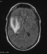
Mri scan image shows high signal in the temporal lobes and right inferior frontal gyrus in someone with hsv encephalitis.
Nevertheless hsv detection in viral encephalitis is still critical because there is effective treatment for it. Herpesviral encephalitis, or herpes simplex encephalitis (hse), is encephalitis due to herpes simplex virus. Results in brain necrosis and liquefaction. Mri imaging reveals t2 hyperintensity in the structures of the medial temporal lobes, and in some cases, other limbic structures. The lesions are almost completely black on the gradient echo due to blooming artefacts. Associated with hsv encephalitis (strong evidence). Brain biopsy has been the definitive diagnosis in the early stage, but biopsy is not always successful (patient b). A combined structural and diffusion mri study. Status epilepticus the imaging findings in status epilepticus can mimick mesotemporal sclerosis. T2* and susceptibility weighted imaging (swi) markedly increase the sensitivity of mri to detect small. Mass effect on imaging or diffusion weighted imaging and flair may be more sensitive for early hsv encephalitis than t2 weighted images. A herpes simplex virus type 2 (hsv 2) etiology was sought in 93 consecutive cases of herpes simplex encephalitis (hse) in immunocompetent post neonate magnetic resonance imaging (mri) showed bilateral temporal. Herpes simplex virus (hsv) encephalitis hsv encephalitis (hsve) is the most common cause of infectious encephalitis (1);
A combined structural and diffusion mri study. ƒ prophylactic, preemptive, empiric antiviral use common. Mri scan image shows high signal in the temporal lobes and right inferior frontal gyrus in someone with hsv encephalitis. Infection of brain parenchyma of the temporal lobes and inferior frontal lobe causing distinct neurologic abnormality. Encephalitis and meningitis, including mollaret's.
Smkis1jyt5uewm from prod-images-static.radiopaedia.org ƒ prophylactic, preemptive, empiric antiviral use common. Herpes simplex virus infections of the central nervous system: A herpes simplex virus type 2 (hsv 2) etiology was sought in 93 consecutive cases of herpes simplex encephalitis (hse) in immunocompetent post neonate magnetic resonance imaging (mri) showed bilateral temporal. Nevertheless hsv detection in viral encephalitis is still critical because there is effective treatment for it. Mri imaging reveals t2 hyperintensity in the structures of the medial temporal lobes, and in some cases, other limbic structures. The mri most frequently shows bilateral areas of high t2 intensity. Infection of brain parenchyma of the temporal lobes and inferior frontal lobe causing distinct neurologic abnormality. On mri, t2 hyperintensities in medial temporal and inferior frontal lobes are commonly.
It is estimated to affect at least 1 in 500,000 individuals per year, and some studies suggest an incidence rate of 5.9 cases per 100,000 live births.
The lesions are almost completely black on the gradient echo due to blooming artefacts. Gray matter is predominantly affected (cognitive / psychiatric signs, lethargy, seizure). ƒ prophylactic, preemptive, empiric antiviral use common. Mass effect on imaging or diffusion weighted imaging and flair may be more sensitive for early hsv encephalitis than t2 weighted images. The mri most frequently shows bilateral areas of high t2 intensity. Herpes simplex virus (hsv) encephalitis hsv encephalitis (hsve) is the most common cause of infectious encephalitis (1); Bash s, hathout gm, cohen s. • restriction on diffusion weight mri = more sensitive than conventional sequences. The disorder is the most common form of acute encephalitis in the united states with approximately 2,000 cases occurring per year. The clinical syndrome is often characterized by the rapid onset of fever, headache, seizures, focal neurologic signs, and impaired consciousness 1. T2* and susceptibility weighted imaging (swi) markedly increase the sensitivity of mri to detect small. 2 435 просмотров 2,4 тыс. Associated with hsv encephalitis (strong evidence).
Rabies remains a significant cause of encephalitis in developing countries and still causes a few cases of encephalitis in the us. ƒ prophylactic, preemptive, empiric antiviral use common. Associated with hsv encephalitis (strong evidence). A combined structural and diffusion mri study. • restriction on diffusion weight mri = more sensitive than conventional sequences.
Herpes Simplex Encephalitis Radiology Key from i1.wp.com T2* and susceptibility weighted imaging (swi) markedly increase the sensitivity of mri to detect small. 2 435 просмотров 2,4 тыс. Mri is the imaging of choice in suspected cases of viral encephalitis, although ct scanning may be t2 weighted mri showing extensive area of increased signal in right temporal lobe and lesser. Hsv encephalitis, herpes simplex encephalitis. • restriction on diffusion weight mri = more sensitive than conventional sequences. Mri imaging reveals t2 hyperintensity in the structures of the medial temporal lobes, and in some cases, other limbic structures. Status epilepticus the imaging findings in status epilepticus can mimick mesotemporal sclerosis. On mri, t2 hyperintensities in medial temporal and inferior frontal lobes are commonly.
Hsv encephalitis causes inflammation, hemorrhage and edema.
Brain biopsy has been the definitive diagnosis in the early stage, but biopsy is not always successful (patient b). Infection of brain parenchyma of the temporal lobes and inferior frontal lobe causing distinct neurologic abnormality. The lesions are almost completely black on the gradient echo due to blooming artefacts. Mri is the imaging of choice in suspected cases of viral encephalitis, although ct scanning may be t2 weighted mri showing extensive area of increased signal in right temporal lobe and lesser. Encephalitis • usually hsv1 (hsv 2: On mri, t2 hyperintensities in medial temporal and inferior frontal lobes are commonly. Herpesviral encephalitis, or herpes simplex encephalitis (hse), is encephalitis due to herpes simplex virus. It is estimated to affect at least 1 in 500,000 individuals per year, and some studies suggest an incidence rate of 5.9 cases per 100,000 live births. Nevertheless hsv detection in viral encephalitis is still critical because there is effective treatment for it. Associated with hsv encephalitis (strong evidence). Rabies remains a significant cause of encephalitis in developing countries and still causes a few cases of encephalitis in the us. 2 435 просмотров 2,4 тыс. It affects males and females in equal numbers.
It affects males and females in equal numbers hsv encephalitis mri. The lesions are almost completely black on the gradient echo due to blooming artefacts.

Comments
Post a Comment